NEWS
Scientists develop the world's first 3D color X-rays

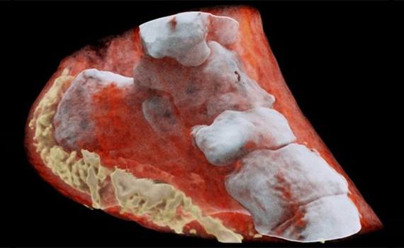
New Zealand scientists have made the world's first three-dimensional color X-ray image of a man using technology that promises to improve the field of medical diagnostics.
A new device, based on a traditional black and white X-ray, uses particle tracking technology developed for the Large Hadron Collider, “Naukatv reports. CERN technology, called Medipix, works as a camera that detects and counts individual subatomic particles when they collide with pixels when the shutter is open. This allows you to use images with high resolution and high contrast.
According to CERN engineers, the images clearly show the difference between bone tissue, muscles and cartilage. Also, the position and size of cancerous tumors are clearly visible. More accurate images help physicians diagnose diseases more accurately.
The technology is already being commercialized by the New Zealand company MARS Bioimaging, which is associated with Otago and Canterbury universities, which participated in the development of the project.


