INSTITUTE OF INFORMATION TECHNOLOGY
The first color images produced by an electron microscope
November 11, 2016 | 02:30 / Interesting information
Electron microscopes are renowned for their ability to peer down into the hidden world of the very small. Trouble is, these tools only produce images in black and white. A new technique that took 15 years to develop finally overcomes this optical limitation, producing the first ever multicolor electron microscope images.
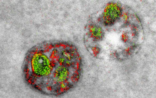
A research team from the University of California, San Diego is the first to create a multicolor electron microscope, allowing for three colors at a time (green, red, and yellow). Technically speaking, the microscope is not producing “true” color images, but rather a false-color visualization of key features found within microscopic objects, such as cells. Importantly, the colors are not “added” after the fact—they’re genuinely indicative of discrete biological components.
Conventional electron microscopes form images by transmitting electron beams through an object, like a biological specimen. This allows for the creation of a detailed monochrome image, but because the microscope is shooting out electrons, and not colored light, there’s a definite absence of color.
To create the colorized scans, the researchers fitted a special detector on a conventional electron microscope. The researchers then selectively “painted” structures such as proteins, membranes, and cells with various “rare earth” metals, including lanthanum, cerium and praseodymium in the form of a chemical solution. When these specimens were scanned by the modified microscope, the stream of electrons lost by the metallic elements were interpreted as color.
© All rights reserved. Citing to www.ict.az is necessary upon using news