NEWS
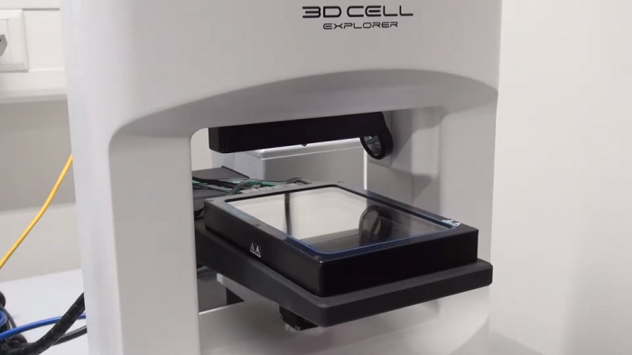
Created a 3D microscope that does not damage the cells in research

Unlike standard methods, the new approach keeps cells alive and healthy. This allows you to track their development and reactions to environmental changes over the long term.
To study a living cell with a microscope, it must be stained. However, standard dyes cause damage and cell death. An alternative technique was presented by researchers from the Swiss Federal Institute of Technology, the work of which tells Engadget.
The team has developed a new three-dimensional microscope CX-A, which allows to examine the internal structures of cells less than 200 nm in size. According to the principle of operation, the device resembles an MRI apparatus: after taking many pictures from different angles, it connects them into a single three-dimensional image. Damage to the cells does not occur.


